
A couple of weeks ago, my kid brother came home from school, dumped his backpack on the kitchen table, and let out a massive sigh. He was holding a sheet of blank paper and a box of colored pencils. “I have to memorize twenty different parts of a cell by tomorrow morning,” he said, sounding completely defeated. “They all look like microscopic blobs, and the names sound like bad sci-fi villains. What even is a Golgi apparatus?”
It brought me right back to my own 9th-grade biology class. I remembered sitting under flickering fluorescent lights, squinting through a cheap school microscope at a slide of onion skin. All I could see was a blurry grid of faint boxes. I didn’t see any “powerhouses,” shipping centers, or recycling bins. It just looked like transparent jelly.
But then my teacher told us something that changed how I looked at biology forever: “Stop trying to memorize a list of dead words. A cell is just a bustling, high-tech city. Every structure inside it is a factory, a power plant, a security gate, or a city hall.”
Suddenly, the dense textbook jargon faded away, and a logical blueprint emerged. If you’re a freshman trying to ace an upcoming unit test, a parent trying to help your teenager study without looking clueless, or just someone who wants a clear, non-boring refresher on how life works at a foundational level, this guide is for you. Let’s open up the microscope and break down cell structure and function using real-world logic that actually sticks.
The Two Big Categories: Prokaryotes vs. Eukaryotes
Before we tour our microscopic city, we need to understand the two main types of cells on Earth. Think of this like the difference between a simple, one-room off-grid cabin and a massive, multi-story smart home.
- Prokaryotes (The Simple Cabin): These are tiny, single-celled organisms like bacteria. They don’t have a separate, walled-off room for their DNA. Everything just floats around together in one open space. They are simple, rugged, and efficient.
- Eukaryotes (The Smart Home): These are more complex cells found in plants, animals, fungi, and you. They have highly specialized, internal “rooms” called organelles (which literally translates to “tiny organs”). Each organelle has its own specific job and its own set of walls.
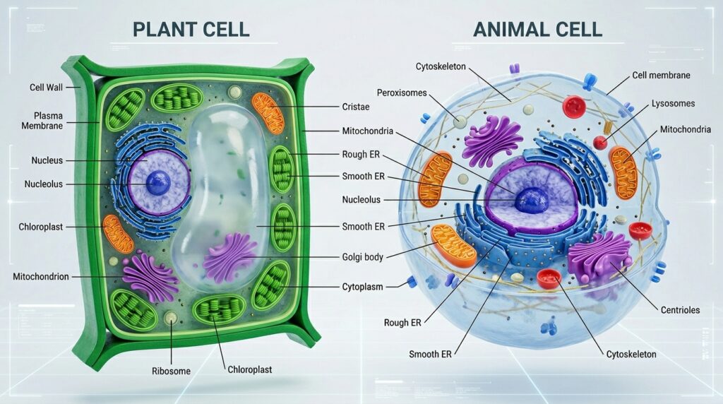
For 9th-grade biology, your main focus is going to be on Eukaryotic cells—specifically, the classic battle between Plant Cells and Animal Cells.
Anatomy of the Cell City: The Core Organelles
Let’s look at a clear visual breakdown of how these structures actually look side-by-side, and then we will examine what each part does for the cell’s survival.
Using our city blueprint metaphor, let’s go building by building through the organelles you’ll see labeled on your diagrams.
1. The Nucleus (City Hall)
The nucleus is the command center of the cell. It holds your DNA, which acts as the master blueprint for absolutely everything the cell constructs.
- The Nucleolus: Nestled deep inside the nucleus, this is like the factory floor inside city hall where the cell builds ribosomes (which we’ll get to in a second).
2. The Cell Membrane (The Border Control & Security Gate)
A flexible, double-layered barrier made of lipids (fats) that wraps around the entire cell. It is “semi-permeable,” meaning it acts like a strict security guard at a gated community. It decides exactly what gets to come in (like water and nutrients) and what gets kicked out (like waste products).
3. The Cytoplasm (The City Terrain)
This is the thick, jelly-like fluid that fills up the empty space inside the cell. Think of it as the air, water, and physical space where all the other buildings and factories are sitting. It keeps everything suspended and cushioned.
4. The Mitochondria (The Power Plant)
You’ve probably seen the internet memes calling this the “powerhouse of the cell.” It’s an accurate title. The mitochondria take the food you eat (glucose) and convert it into a chemical fuel cell called ATP. Without mitochondria, the cell completely runs out of juice and shuts down.
5. The Endoplasmic Reticulum (The Highway System & Factory Complex)
Often just abbreviated as the ER, this is a massive network of folded membranes twisting out from the nucleus. It is the cell’s industrial manufacturing zone, split into two sectors:
- Rough ER: Covered in tiny dots called ribosomes. This makes it look bumpy or “rough.” This is where proteins are actively built.
- Smooth ER: Has no ribosomes. Its job is to manufacture lipids (fats) and break down toxins or poisons.
6. Ribosomes (The Construction Workers)
These are tiny, microscopic machines that read instructions sent out from the nucleus and stitch together proteins. They are the manual laborers of the cellular world. You’ll find them stuck to the Rough ER or floating freely through the cytoplasm.
7. The Golgi Apparatus (The Post Office / Shipping Center)
Once the ribosomes and Rough ER build proteins, those proteins get sent to the Golgi apparatus. The Golgi looks like a stack of deflated pancakes. It inspects the proteins, customizes them with molecular tags, packs them into tiny bubble-like shipping boxes called vesicles, and mails them to wherever they need to go inside or outside the cell.
8. Lysosomes & Peroxisomes (The Waste Management & Recycling Crew)
These are small sacs filled with digestive enzymes. Think of them as the garbage trucks and recycling plants. They track down worn-out organelles, broken proteins, or invading bacteria, break them apart into raw materials, and clean up the cell city.
Plant vs. Animal Cells: Spotting the Key Differences
This is the most heavily tested concept in freshman biology. Teachers love to put a diagram on a quiz and ask you to identify if it’s a plant or an animal cell.
If you look back at our diagram above, you’ll see that while they share almost all of the same machinery, plant cells have three exclusive features that animal cells completely lack.
| Feature / Organelle | Plant Cells | Animal Cells | Why It Matters |
| Shape | Rigid, boxy, rectangular | Round, flexible, irregular | Plants need structure to stand upright; animals have skeletons for support instead. |
| Cell Wall | Yes | No | A tough outer layer made of cellulose that sits outside the membrane for extra protection. |
| Chloroplasts | Yes | No | Green solar panels that convert sunlight into sugar via photosynthesis. |
| Vacuole Size | One Massive Central Vacuole | Multiple tiny vacuoles | Plants use the huge water pressure inside their central vacuole to keep their stems crisp and straight. |
Step-by-Step Guide to Identifying a Cell on a Test
When you face an unlabeled cell diagram on an exam, don’t guess. Use this simple, elimination-style check sequence to identify what you’re looking at within seconds.
1.Look for a Nucleus:Step 1.
Find the large, central circle. Is there a clear, bounded nucleus holding the DNA?
- No: It’s a Prokaryotic cell (like a bacterium). Stop here.
- Yes: It’s a Eukaryotic cell. Proceed to Step 2.
2.Check the Outer Border:Step 2.
Look at the outer edge of the cell.
- Is it a single, thin, round layer? It’s an Animal cell.
- Is there a thick, distinct secondary frame enclosing a rectangular shape? It’s a Plant cell.
3.Scan for Solar Panels (Chloroplasts):Step 3.
Look inside the cytoplasm for oval-shaped structures containing little stacks of discs (thylakoids) that look like piles of green coins. If you see them, you are definitely looking at a Plant cell.
4.Examine the Water Tank (Vacuole):Step 4.
Look at the center of the cell. If there is a massive, clear reservoir taking up over 50% of the entire cell’s internal volume, it’s a Plant cell using water pressure to stay rigid.
Classic Mistakes to Avoid
When grading biology quizzes or studying with flashcards, watch out for these common mix-ups:
- Confusing Cell Wall with Cell Membrane: Plant cells do not skip the cell membrane. They have both. The cell membrane is the inner layer controlling what passes through; the cell wall is the thick, rigid exterior scaffolding wrapped around it.
- Thinking Plant Cells Don’t Have Mitochondria: This is a massive trapdoor. Many students assume that because plants have chloroplasts for solar energy, they don’t need mitochondria. That is wrong! The chloroplast builds the food (glucose), but the plant still needs mitochondria to break that food down into usable energy (ATP). Plants have both organelles.
- Mixing up the ER and the Golgi: On messy black-and-white test diagrams, these two look very similar because they’re both folded lines. Look at the location: the ER is always physically attached to the nucleus, while the Golgi apparatus floats further away toward the edge of the cell.
Free Digital Tools to Master This Visually
If staring at flat, 2D diagrams in an old textbook is making you cross-eyed, close the book and try out these interactive resources:
- BioDigital Human / Cell Models: An incredible, free web platform that lets you explore fully immersive 3D interactive models of cells. You can rotate the cell, slice it in half, zoom in on a single ribosome, and click on parts to see them animate in real-time.
- Bioman Biology: A great educational gaming site filled with arcade-style games centered around cell structure. Games like “Cell Defense” teach you how the cell membrane works by letting you actively guard the gate against incoming molecules.
Final Thoughts
Biology looks incredibly intimidating when you approach it as a giant word search puzzle of Latin roots and complex labels. But beneath the surface, life runs on pure organization. Every living cell is just a highly synchronized microscopic community working together to keep the power on, the borders secure, and the blueprints safe.
Keep that city map in your head, practice sketching out the borders a couple of times on scratch paper, and those test questions won’t stand a chance.